
MODI II Keratoconus Screening Case

OU Topography Report:
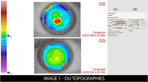
Tangential maps above show substantial differences between eyes.
The OS map shows a large amount of asymmetry in curvature with inferior steepening.
The OD map shows some astigmatism – mildly asymmetrically.
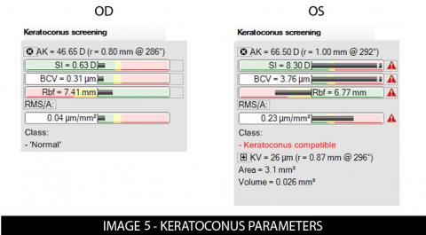
The parameters shown to the right present the keratoconus screening parameters for each eye.
- The OS parameters identify Outside Normal Limits with the explanation mark
- The software identifies the left eye as Keratoconus Compatible
- The OD parameters all fall within the Normal range
4UP Summary OS:
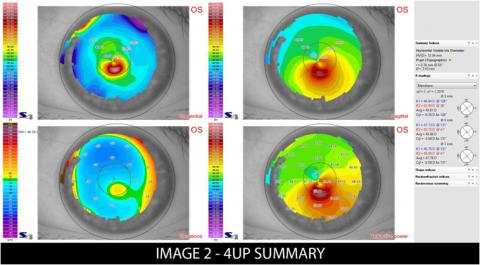
The top left map shows local curvature and top right map shows global curvature.
Inferior steepening consistent with keratoconus is clearly displayed.
The elevation map (bottom left) show inferior vaulting in the corneal elevation, consistent with the classic presentation of a keratoconus .
Keratoconus Summary:
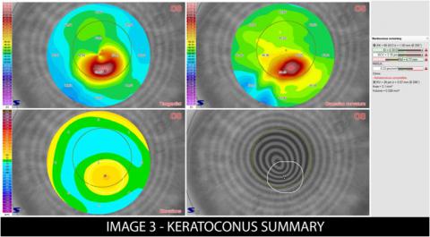
Specializes maps for keratoconus screening and diagnosis , displayed in summary.
All summary report maps are shown with a radius of 3mm
The top left map shows the asymmetric astigmatism of the Tangential (local) curvature.
At the top right a Gaussian curvature map is displayed. This presentation map is useful as it removes normal astigmatism from the map but preserves the ectasia.
The bottom left is a specialised Elevation Map that uses a toric ellipsoid as the reference plane. This better approximates the corneal shape so the elevated cone is clearly localised and quantified.
Bottom right map displays the area of suspect curvature traced onto the eye image for easier localization.
Keratoconus Parameters:
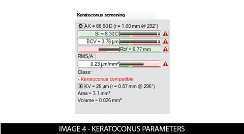
The MODI uses a normative database with the following corneal parameters to identify patients with Keratoconus:
Visually, they are presented in a bar showing how that parameter compares to normal
Keratoconus Parameters (con’t):
The MODI software then uses a neural network to process the parameters and make a determination about the patient’s condition
When the topographic context appears compatible with keratoconus or indicates a suspicion of keratoconus, a series of morphological indicators are presented for evaluation:
In the case presented here, the OD parameters are normal and all the OS parameters indicate keratoconus . The patient is flagged as Keratoconus Compatible with additional parameters displayed.